2 Institute of Human Genetics, Schwabachanlage 10, D-91054 Erlangen, Germany
3 FRIGE’s-Institute of Human Genetics, Cytogenetic and Molecular Cytogenetic Dept Indian FRIGE, Institute of Human Genetics, Jodhpur Road, Satellite, Ahmedabad 380 015, Gujarat, India
4 Samodzielna Pracownia Cytogenetyki, Centrum Onkologii-Instytut, ul.W.K.Roentgena 5, 02-781 Warszawa, Poland
5 Department of Genetic and Laboratory of Cytogenetics, State University, Yerevan, Armenia
Abstract
The impact of chromosome architecture in the formation of chromosome aberrations is a recent finding of interphase directed molecular cytogenetic studies. There evidence was provided that disease specific chromosomal translocations could be due to tissue specific genomic organization. In a recent small pilot study using three-dimensional interphase fluorescence in situ hybridization, we showed that there might be a specific chromosome positioning in myeloid bone marrow cells, i.e. a co-localization of chromosomes 8 and 21. Here we could substantiate this finding in overall 21 studied cases with acute myeloid leukemia (AML) that there is even a co-localization of the genes AML1 and ETO. This finding led to the suggestion that a specific interphase architecture of myeloid bone marrow cells might promote the typical t(8;21)(q22;q22) leading to AML-M2.
Citation: Othman M, Lier A, Junker S, Kempf P, Dorka F, Gebhart E et al. Does positioning of chromosomes 8 and 21 in interphase drive t(8;21) in acute myelogenous leukemia? BioDiscovery 2012; 4: 2; DOI: 10.7750/BioDiscovery.2012.4.2
Copyright: © 2012 Othnam et al. This is an open-access article distributed under the terms of the Creative Commons Attribution License, which permits unrestricted use, provided the original authors and source are credited.
Received: September 28, 2012; Accepted: October 16, 2012; Available online /Published: October 23, 2012
Keywords: interphase chromosome (genome) organization, formation of chromosome aberrations, fluorescence in situ hybridization (FISH), chromosome 8, chromosome 21
*Corresponding Author: Thomas Liehr, e-mail: i8lith@mti.uni-jena.de
Conflict of Interests: No potential conflict of interest was disclosed by any of the authors.
Introduction
Analysis of the location of chromosomes and genes in a number of cell types and tissues has revealed that “genomic elements” (i.e. here chromosomes) occupy preferential positions within the nucleus which are called ‘chromosome territories’ [1-3]. Chromosome size and density of the genes within a chromosome are discussed to have an impact on the nuclear position of chromosomes [4]. Furthermore, non-random positioning in interphase nuclei is known to be of importance for genomic stability and formation of chromosome aberrations. Tissue specificity of chromosomal translocations could be due to tissue specific genome organization [5-6], and a positive correlation between spatial proximity of chromosomes/ genes in interphase nuclei and translocation frequencies was shown [5-10]. Three-dimensional (3D) fluorescence in situ hybridization (FISH) analysis became a major tool for studying this higher order chromatin organization in the cell nucleus [4; 11-15].
Trisomy 8, the most frequently occurring numerical chromosome aberration in acute myeloid leukemia (AML) and myelodysplastic syndromes (MDS), can be associated with other karyotypic abnormalities, or occur as sole abnormality. Trisomy 8 is also a marker for progression in chronic myelogeneous leukemia (CML). A variety of hematological diseases are connected with trisomy 8, indicating a non-specific role in leukemia pathogenesis. The prognostic impact of trisomy 8 as the sole change in AML and MDS is discussed controversial in the literature [16-19]. However, another frequent cytogenetic abnormality involving chromosome 8, the reciprocal translocation t(8;21) usually correlates with AML-M2 and indicate a good prognosis [20]. The AML1-ETO (also RUNX1/MTG8) fusion oncoprotein, generated by the t(8;21) chromosomal translocation, is causally involved in nearly 15% of AML cases. AML1/ ETO consists of the N-terminal DNA-binding domain of AML1, a transcription factor essential for definitive hematopoiesis, and almost all of ETO, a protein thought to function as a co-repressor for a variety of transcription factors [21].
In a previous study the (relative) 3D position of chromosomes 8 and 21 to each other was studied in interphase nuclei of AML cases with trisomy 8 [11]. In the present study we enlarged the number of cases and analyzed relative position of not only chromosomes 8 and 21 [11] but also the genes suggested to be involved ETO and AML1. Bone marrow (BM) of AML, MDS and CML cases with trisomy 8 were studied in comparison with BM of six AML-M2 cases in remission, BM of two control cases with autoimmune thrombocytopenia and PBL of four healthy donors, each with normal karyotype. The well established approach of interphase chromosome-specific multicolor banding (ICS-MCB) [15], partial chromosome paining (PCP) probes and LSI AML1/ETO Dual Color, Dual Fusion Translocation Probe (Vysis) combined with suspension FISH (S-FISH) [14] were chosen for this study.
Materials and methods
The studied patients are listed in Table 1.
![Table 1. Overview of the 21 cases with and without trisomy 8, studied material and karyotypes; AML-M2 cases had originally a t(8;21). Cases with asterisks were previously published in Manvelyan et al. [11]. Abbreviation: mMCB = multitude multicolor banding [25]. Table 1](https://web.archive.org/web/20160322133437im_/http://biodiscoveryjournal.co.uk/Archive/Media/A16Table1-550.jpg)
Multicolor banding (MCB) probe sets for chromosomes 8 and 21 were applied in suspension- FISH (S-FISH) as previously reported [11]. Images of 3D-preserved interphase nuclei were captured on a Zeiss Axioplan microscope and analyzed by Cell-P (Olympus) software. In the same way, the LSI AML1/ETO Dual Color – Dual Fusion Translocation Probe (Vysis) was used and analyzed.
For the 3D-evaluation, position and distance of homologous chromosomes/ signals were determined. The interphase nucleus was divided into two spheres, i.e. periphery (P) and center (C); 50% of the nucleus radius was defined as ‘center’. Thus, analyzed chromosomes could be allocated either as C or P. The relative positions of the studied chromosomes to each other were recorded as ‘close together’ (t), ‘near by each other’ (n) or ‘on the opposite sides of the nucleus’ (o) for two homologue chromosomes. In cells with three chromosomes 8 this nomenclature was combined to ‘o-n’, ‘o-t’ or ‘t-n’ – for examples see [11].
Statistical analysis was performed using Student’s t-test, One Way ANOVA (Analysis of Variance) and Holm-Sidak method. Statistical significance was defined as p<0.05.
Results and discussion
In the present study we found that chromosome 8 is predominantly positioned in the periphery (P) of interphase nuclei. The position of chromosome 8 in BM cells and peripheral blood-lymphocytes is in concordance with the data of our previous study determined in haploid human sperm [4]. The additional chromosome in trisomy 8 was located in periphery rather than central. If this is general behavior of additional chromosomes present cannot be answered yet. According to the present study, homologue chromosomes 8 are located primarily in close proximity, i.e. close together (t).
Observed position of chromosome 21 was in concordance with the literature [22-23]. In all here studied diploid cases, homologue chromosomes 21 behaved as postulated for acrocentrics and co-localized to each other in a more central position. Previously, it was postulated [22-23] that their co-localization is caused by the nucleolar organizer regions on their short arms, and that the nucleolus is located in the inner nuclear space.
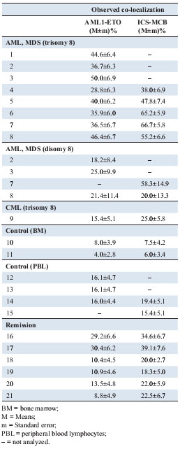
Correlation between spatial proximity of chromosomes/ genes in interphase nuclei and translocation frequencies was shown before and chromosomes located in proximity underwent translocation events more frequently than distantly located ones [6; 10; 12]. To test this hypothesis for the reciprocal translocation t(8;21) usually correlated with AML, here a 3D analysis for co-localization of chromosomes 8 and 21 was done (Table 2).

As reported [11] a random co-localization was compared to the observed co-localization rate of one chromosome 8 and 21, each. This was done based on interphase ICS-MCB applied in S-FISH and using locus-specific probes for the AML1/ETO translocation (Figure 1). A significant enhanced co-localization rate was found in all studied trisomy 8 AML-cases (Table 2), with exception of trisomic cells of case 4 and disomic cells of cases 3 and 8, compared to controls. In all other cases (normal controls, CML and AML-M2 in remission) practically no co-localization of one chromosome 8 and 21 was observed, with exception of case 17.

Generally, in trisomy 8 cells there was a significant co-localization of the locus-specific probes AML1 and ETO in 7 out of 8 cases, while by ICS-MCB and looking at whole chromosomes 8 and 21 such a correlation was only observable in 2 out of five cases. Even in two out of four of these cases, the cells with disomy 8 showed a significant co-localization of one chromosome 8 and 21 including AML1 and ETO. Neither in the one CML- case with trisomy 8 nor in stimulated peripheral blood T-lymphocytes was this co-localization found; also not in BM of patients with autoimmune thrombocytopenia. Finally, in one out of the six cases with AML2 in remission a co-localization of AML1 and ETO could be proven. This inconsistency of chromosome 8 and 21 co-localization might also point towards new entities of AML2 distinguishable only by 3D-FISH analysis. Also, in cases with trisomy 8 and AML1-ETO fusion, t(8;21) (q22;q22) might have to be considered as secondary rather than primary event. Still, at present it is not clear if a co-localization of chromosomes 8 and 21 promotes a translocation between the two chromosomes in AML-M2 or even in AML-cases with trisomy 8.
Overall, further studies in AML are necessary for delineation of interphase architecture in this cell type as in cancer in general. At present, as supported by recent comparable findings in thyroid cancer, a clinical impact of 3D-chromosome positioning on malignancies becomes more and more likely [24].
Acknowledgments
This work was partly supported by the Stefan-Morsch-Stiftung, the DAAD and the DFG (LI 820/21-1 and LI 820/24-1).
References
- Cremer T, Cremer M: Chromosome Territories: Cold Spring Harb. Perspect Biol 2010; 2:a003889.
REFERENCE LINK - Takizawa T, Meaburn KJ, Misteli T: The Meaning of Gene Positioning. Cell 2008; 135: 9-13.
REFERENCE LINK - Lemke J, Claussen J, Michel S, Chudoba I, M?hlig P, Westermann M, et al.: The DNA-based structure of human chromosome 5 in interphase. Am J Hum Genet 2002; 71: 1051-1059.
- Manvelyan M, Hunstig F, Bhatt S, Mrasek K, Pellestor F, Weise A, et al.: Chromosome distribution in human sperm – a 3D multicolor banding study. Mol Cytogenet 2008; 1: 25.
REFERENCE LINK - Meaburn KJ, Misteli T, Soutoglou E: Spatial genome organization in the formation of chromosomal translocations. Semin Cancer Biol 2007; 17: 80-90.
REFERENCE LINK - Brianna Caddle L, Grant JL, Szatkiewicz J, van Hase J, Shirley BJ, Bewersdorf J, et al.: Chromosome neighborhood composition determines translocation outcomes after exposure to high-dose radiation in primary cells. Chromosome Res 2007; 15: 1061-1073.
REFERENCE LINK - Branco MR, Pombo A: Intermingling of chromosome territories in interphase suggests role in translocations and transcription dependent associations. PLoS Biol 2006; 4: E138.
REFERENCE LINK - Grasser F, Neusser M, Fiegler H, Thormeyer T, Cremer M, Carter NP, et al.: Replication-timing correlated spatial chromatin arrangements in cancer and in primate interphase nuclei. J Cell Sci 2008; 121: 1876-1886.
REFERENCE LINK - Bickmore WA, Teague P: Influences of chromosome size, gene density and nuclear position on the frequency of constitutional translocations in the human population. Chromosome Res 2002; 10: 707-715.
REFERENCE LINK - Roix JJ, McQueen PG, Munson PJ, Parada LA, Misteli T: Spatial proximity of translocation-prone gene loci in human lymphomas. Nat Genet 2003; 34: 287-291.
REFERENCE LINK - Manvelyan M, Kempf P, Weise A, Mrasek K, Heller A, Lier A, et al.: Preferred co-localization of chromosome 8 and 21 in myeloid bone marrow cells detected by three dimensional molecular cytogenetics. Int J Mol Med 2009; 24: 335-341.
- Cremer M, von Hase J, Volm T, Brero A, Kreth G, Walter J, et al.: Non-random radial higherorder chromatin arrangements in nuclei of diploid human cells. Chromosome Res 2001; 9: 541-567.
REFERENCE LINK - Solovei I, Cavallo A, Schermelleh L, Jaunin F, Scasselati C, Cmarko D, et al.: Spatial preservation of nuclear chromatin architecture during three-dimensional fluorescence in situ hybridization (3D-FISH). Exp Cell Res 2002; 276: 10-23.
REFERENCE LINK - Steinhaeuser U, Starke H, Nietzel A, Lindenau J, Ullmann P, Claussen U, et al.: Suspension (S)-FISH, a new technique for interphase nuclei. J Histochem Cytochem 2002; 50: 1697-1698.
REFERENCE LINK - Iourov IY, Liehr T, Vorsanova SG, Yurov YB: Interphase chromosome-specific multicolor banding (ICS-MCB): a new tool for analysis of interphase chromosomes in their integrity. Biomol Eng 2007; 24: 415-417.
REFERENCE LINK - Paulsson K, Johansson B: Trisomy 8 as the sole chromosomal aberration in acute myeloid leukemia and myelodysplastic syndromes. Pathol Biol (Paris) 2007; 55: 37-48.
REFERENCE LINK - Heller A, Brecevic L, Glaser M, Loncarevic I, Gebhart E, Claussen U, et al.: Trisomy 8 as the sole chromosomal aberration in myelocytic malignancies: a comprehensive molecular cytogenetic analysis reveals no cryptic aberrations. Cancer Genet Cytogenet 2003; 146: 81-83.
REFERENCE LINK - Makishima H, Rataul M, Gondek LP, Huh J, Cook JR, Theil KS, et al.: FISH and SNP-A karyotyping in myelodysplastic syndromes: improving cytogenetic detection of del(5q), monosomy 7, del(7q), trisomy 8 and del(20q). Leuk Res 2010; 34: 447-453.
REFERENCE LINK - Paulsson K, Heidenblad M, Str?mbeck B, Staaf J, J?nsson G, Borg A, et al.: High-resolution genome-wide array-based comparative genome hybridization reveals cryptic chromosome changes in AML and MDS cases with trisomy 8 as the sole cytogenetic aberration. Leukemia 2006; 20: 840-846.
- Lai YY, Qiu JY, Jiang B, Lu XJ, Huang XJ, Zhang Y, et al.: Characteristics and prognostic factors of acute myeloid leukemia with t(8,21)(q22,q22). Zhongguo Shi Yan Xue Ye Xue Za Zhi 2005; 13: 733-740.
- Petrie K, Zelent A: AML1/ETO: A promiscuous fusion oncoprotein. Blood 2007; 109: 409-410.
REFERENCE LINK - Sun HB, Shen J, Yokota H: Size-dependent positioning of human chromosomes in interphase nuclei. Biophys J 2000; 79: 184-190.
REFERENCE LINK - Bolzer A, Kreth G, Solovei I, Koehler D, Saracoglu K, Fauth C, et al.: Three-dimensional maps of all chromosomes in human male fibroblast nuclei and prometaphase rosettes. PLoS Biol 2005; 3: E157.
REFERENCE LINK - Gandhi MS, Stringer JR, Nikiforova MN, Medvedovic M, Nikiforov YE: Gene position within chromosome territories correlates with their involvement in distinct rearrangement types in thyroid cancer cells. Genes Chr Cancer 2009; 48: 222-228.
REFERENCE LINK - Weise A, Heller A, Starke H, Mrasek K, Kuechler A, Pool-Zobel BL, et al.: Multitude multicolor chromosome banding (mMCB) – a comprehensive one-step multicolor FISH banding method. Cytogenet Genome Res 2003; 103: 34-39.
REFERENCE LINK


